Research Article
Experimental ‘hindbrain related’ syringomyelia: some mechanisms of spinal cord damage

Sergey N Larionov1,3*, Sorokovikov VA1,2 and Rudakova AV3
1Scientific Center of Reconstructive and Restorative Surgery, RAMS, Siberian Branch, Russia
2Department of Traumatology, Orthopedics and Neurosurgery, Irkutsk State Medical Academy of Continuous Education, Irkutsk, Russia
3Department of Child’s Neurosurgery, Regional Child’s hospital, Irkutsk, Russia
*Address for Correspondence: Sergey N Larionov, Scientific Center of Reconstructive and Restorative Surgery, RAMS, Siberian Branch, Department of Child’s Neurosurgery, Regional Child’s hospital, Irkutsk, Russia, Email: [email protected]
Dates: Submitted: 03 August 2017; Approved: 05 October 2017; Published: 06 October 2017
How to cite this article: Larionov SN, Sorokovikov VA, Rudakova AV. Experimental ‘hindbrain related’ syringomyelia: some mechanisms of spinal cord damage. J Neurosci Neurol Disord. 2017; 1: 033-038. DOI: 10.29328/journal.jnnd.1001006
Copyright License: © 2017 Larionov SN, et al. This is an open access article distributed under the Creative Commons Attribution License, which permits unrestricted use, distribution, and reproduction in any medium, provided the original work is properly cited.
Abstract
Syringomyelia in combination with inherent or acquired hindbrain abnormalities is the non seldom and at the same time controversial issue.
Purpose: The etiology and pathogenesis creates a lot of discussion.
Methods: Experimental syringomyelia was induced in 20 anesthetized rabbits by injecting 0.5 ml of 25% kaolin suspension into the cisterna magna. Six rabbits with puncture and injection sterile saline NaCl were used as a control. The animals were sacrificed 1, 2, 4 and 6 months after the kaolin injection. Four hydrocephalus rabbits were sacrificed in 17 hours after the puncture of lateral ventricle with injection of solution of colloidal gold labeled human albuminum. The sections of the brain and spinal cord were stained with hematoxylin and eosin by Nissle and Marchi methods and with immunogold technique. Retropharyngeal lymph nodes of the animals were examined by electron microscopy.
Conclusion: Our observation showed that water hammer effect and internal destruction of the spinal cord may lead to continuous antigen stimulation of regional lymph nodes and play an important role in pathogenesis of experimental syringomyelia.
Introduction
Numerous experimental observation and clinical researches demonstrated that the majority of the forms of syringomyelia are found in combination with inherent or acquired hindbrain abnormalities [1,2]. Although hydrodynamic disorders play an important role in the pathogenesis, these forms of syringomyelia, several aspects of this disease are difficult to explain only on the basis of hydrodynamics. Different theories of pathogenesis of syringomyelia were proposed such as “cranio-spinal pressure dissociation”, transmedullary passage of CSF, impaired venous drainage [2,3]. However, the precise pathogenesis of “hindbrain related” syringomyelia is unknown. There are some references of possible role antibodies in the development of syringomyelia. Researches in this field are of great interest. It is undoubted, that experimental studies can be used in the explanation of the nature of this important group of neurological disorders, as we do not have possibilities to study directly the human central nervous system.
The aim of the present experimental study was to determine a possible role of lymphatic drainage of the CSF and antigens stimulation of regional lymph nodes in induction of demyelinating lesions for “hindbrain related” syringomyelia.
Matherial and Methods
A study was approved by the Ethical Committee of Scientific Center of Reconstructive and Restorative Surgery. In this study we used 26 New Zeland white rabbits, each weighing 3500 ± 350 g, and the animals were allocated into two groups. Prior the operation all animals endured intramuscular cephazolin sodium injection (20 mg/kg). All procedures were performed while the rabbits were under pentobarbital anesthesia.
Experimental syringomyelia was induced in 20 anesthetized rabbits by injecting 0.5 ml of 25% kaolin suspension into the cisterna magna. In addition, 6 rabbits with puncture and injection sterile saline NaCl were used as a control.
The animals were sacrificed 1, 2, 4 and 6 months (5 animals per period) after the kaolin injection. Four hydrocephalus rabbits (two animals with 4 and two with 6 months of experiment) were sacrificed in 17 hours after the puncture of lateral ventricle with injection of solution of colloidal gold labeled human albumin (0.5 mg HAuC14 in 0.2 ml sterile saline). For sacrificing the animals were anesthetized by intravenous pentobarbital (50 mg/kg). Their descending aorta was clamped, and they were perfused via transcardiac path with 4% paraformaldehyde in 0.12 M phosphate buffer and 0.02 mM calcium chloride at 37 °C. After ventricular injection of gold labeling albumin retropharyngeal lymph nodes of the animals were examined by electron microscopy. The sections of the brain and spinal cord were stained with hematoxylin and eosin by Nissle and Marchi methods and with immunogold technique. The preparations were examined under a light microscope with magnification ×40, 100, 200 and 400. Meningeal fibrosis was examined according to the following scheme designed by He et al. The fibroblast cell density was calculated in each field by the median fibroblast cell count.
Statistical Analysis
Data analysis was performed using SPSS for Windows, version 11.5 [SPSS Inc., Chicago, IL, USA]. The Shapiro-Wilk test was used to determine if the distributions of continuous variables were normally distributed. The Bartlett test was used to evaluate the homogeneity of the variances. Continuous and ordinal variables were shown as medians (min-max). A nonparametric Kruskal-Wallis test was used to determine the statistical significance of the pachymeningeal fibrosis extension, inflammation, intramedullary cyst formation and dilatation of the ventricular system among groups. The differences in the median values among the groups were compared using a Mann-Whitney test. We used the Likelihood Ratio test to determine whether the differences in nominal data were statistically significant. When the p values from the Likelihood Ratio test statistics were statistically significant, we employed Wilcoxon’s exact test to determine which group differed from which other groups. A p value less than 0.05 was considered statistically significant.
Results
The use of the experimental model of kaolin induced syringomyelia showed in the rabbits the following disturbances in CNS. Intramedullary cavities similar to syringomyelia appeared in the cervical, thoracic and lumbar cord (Figure 1). During the early stage of the experiment (2-4 months) inflammatory infiltrates were found around vessels in the meninges, and rarely find in white matter. Sometimes inflammations resulted in honeycombs-like destructions of nervous tissue (Figure 2).
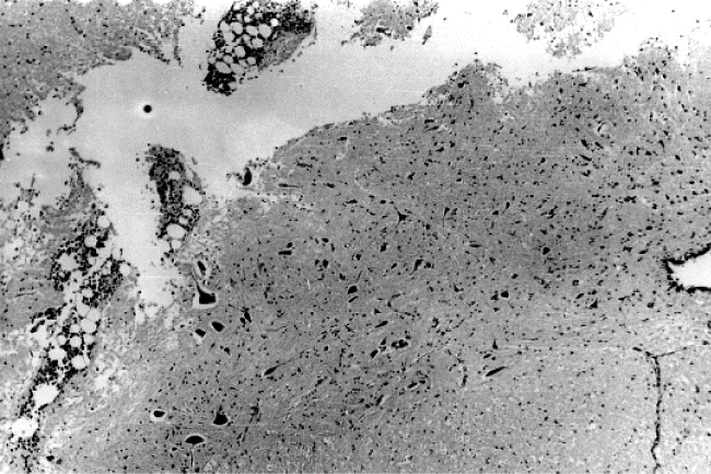
Figure 1: Photomicrographs stained with H & E. A: Photomicrograph of a section of a middle cervical cord demonstrating intramedullary cavity formation in the unilateral posterior column. Original magnification 100.
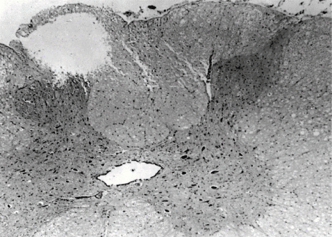
Figure 2: Photomicrographs, of the honeycombs-like destructions spinal cord parenchyma adjacent to the central canal, and infiltration macrophages. Original magnification 100.
In all the animals there was found hydrocephalus but with some variation in the degree of dilatation of the lateral ventricles and of the central lumen of the olfactory bulbs (Figures 3-7).
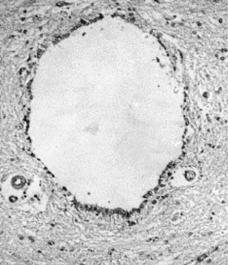
Figure 3: Photomicrographs stained with H & E. A: Mild hydrocephalus, view shows dilated ventricular lumen of the olfactory bulb. Hemathoxylin and eosin. Original magnification 100.
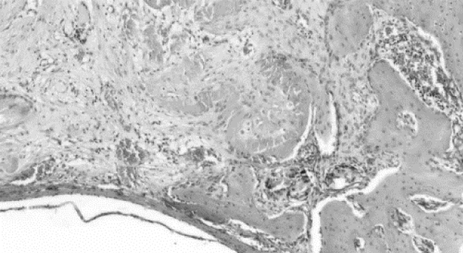
Figure 4: Grade 3fibrosis as observed on the 40th day. Photomicrographs stained with H & E.
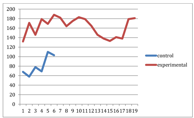
Figure 5: Graph of comparison of fibroblast cell density in the control and experimental groups.
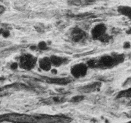
Figure 6: Myelin degradation products in white matter. An oblique section of brain. Marchi method. Original magnification 100.
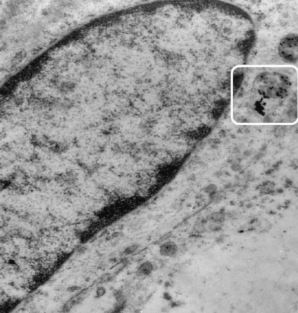
Figure 7: Retropharingeal lymph node of the rabbit afther intraventricular injection of albumin-gold suspension. Gold particles in lysosomas of reticular cells of lateral sinus. Mag.700.
There was significant concordance on all parameters between 2 observers (k coefficient 0.684, 0.712 and 0.702 for pachymeningial fibrosis, intramedullary cyst formation and dilatation of the ventricular system). All of these values were statistically significant (p<0.001).
Fibroblast cell density and meningeal fibrosis were to be less in the control group than in the experimental group (p=0.003, and p=0.005, respectively); these differences were statistically significant also (Table 1).
| Table 1: Comparison of meningeal fibrosis by grade number (%) | |||
| Group | Grade 1 | Grade 2 | Grade 3 |
| Experiment | - (0) | 7 (35.0%) | 13 (65.0%) |
| Control | 4 (66.7) | 2 (33.3%) | – |
After 6 months the experiment of cyst formation, demyelinated plagues in combination with swollen astrocytes were observed. Ultrastructural examination of retropharyngeal lymph nodes of the animals after intraventricular injection of gold labeled albumin revealed gold particles in lysosomas of reticular cells in lateral sinus.
Discussion
It is generally accepted that chronic pachymeningitis of the hindbrain as well as hindbrain anomaly leads to CSF circulation disorders on the level of fourth ventricle outlets. It is an initial mechanisms of dilatation of the ventricular system and cavity formation within the spinal cord. Although hydrodynamic disorders play an important role in the pathogenesis of hydrocephalus and “hindbrain related” syringomyelia, several aspects of disease are difficult to explain only on the basis of hydrodynamics [1,2]. Moreover, indirect evidences show that immune reactions against CNS antigens may play an additional role in the demyelination for “hindbrain related” syringomyelia. However, we cannot explain whether Ig synthesis take place in syringomyelia patients. Blood-brain barrier (BBB) damage is probably an important factor in the problem of determining whether Ig was synthesized in CNS or entered the brain as a result of BBB changes.
Obviously, that the immunological disorders in syringomyelia are of secondary character. Therefore, the following question arises: Why does selective sensibilization against CNS tissue occur. According to the presented data, it is clear if at some moment of time a mitogenic dose of neuroantigen reaches draining lymph nodes, then the proliferation of specific neuroantigen-sensitized cells occur. This mechanisms allows auto reactive clones of lymphoid cells to act directly against CNS antigens. Lymphatic drainage of the CSF has been well documented in a number of mammalian species [4,5]. Then, by highly directionalised pathways, CSF drains to the olfactory bulbs and through arachnoid channels to connect with lymphatic vessels in the nasal mucosa and deep cervical lymph nodes. Our observation on albumin-gold complex reaching to retropharyngeal lymph nodes after ventricular injection of tracer showed that water hammer effect and internal destruction of the spinal cord may lead to continuous antigen stimulation of regional lymph nodes. Anatomical pathways for lymphatic drainage of interstitial fluid, analogous to those in the animals, are present in the human brain [6,7]. It testifies that lymphatic system of head and neck take part in CSF resorbtion. Therefore, it is possible that in humans the continuous antigen stimulation of regional lymph nodes may lead to the induction of chronic demyelination as it happens in the experimental animals. Cervical lymphatic lymph nodes play significant roles in the induction of immunological tolerance and of adaptive immunological responses. The identification and characterization of antigens involved in the pathogenesis of syringomyelia on animals model of disease may lead to a greater understanding of thus pathology, to increase possibilities of therapy for diseases such as syringomyelia.
References
- Levine DN. The pathogenesis of syringomyelia associated with lesions at the foramen magnum: a critical review of existing theories and a proposal of a new hypothesis. J Neurol Sci. 2004; 220: 3-21. Ref.: https://goo.gl/o6ZSse
- Williams B. Simultaneous cerebral and spinal fluid pressure recordings. Cerebrospinal dissociation with lesions at the foramen magnum. Acta Neurochir. 1981; 59: 123-142. Ref.: https://goo.gl/9W2xbr
- Ball MJ, Dayan AD. Pathogenesis of syringomyelia. Lancet. 1972; 2: 799-801. Ref.: https://goo.gl/x98d8F
- Cserr HF, Harling-Berg CJ, Knopf PM. Drainage of brain extracellular fluid into blood and deep cervical lymph and its immunological significance. Brain Pathol. 1992; 2: 269-276. Ref.: https://goo.gl/fvbiVB
- Bakker EN, Bacskai BJ, Arbel-Ornath M, Aldea R, Bedussi B, et.al. Lymphatic clearance of the brain: perivascular, paravascular and significance for neurodegenerative diseases. Cellular and Molecular Neurobiology. 2016; 36: 181-194. Ref.: https://goo.gl/mTGQXT
- Kida S, Pantazis A, Weller RO. CSF drains directly from the subaracnoid space into nasal lymphatics in rat. Anatomy, histology and immunological significance. Neuropath Appl Neurobiol. 1993; 19: 480-488. Ref.: https://goo.gl/XTtQFd
- Weller RO, Djunda E, Yow HY, Carare RO. Lymphatic drainage of the brai and pathophysiology of neurological disease. Acta Neuropathol. 2009; 117: 1-14. Ref.: https://goo.gl/oZ7XRp