Figure 1
Primary intracranial Hodgkin’s lymphoma after a blunt trauma: A case report
Luca Riccioni*, Anna Maria Cremonini and Manlio Gessaroli
Published: 15 December, 2020 | Volume 4 - Issue 2 | Pages: 079-083
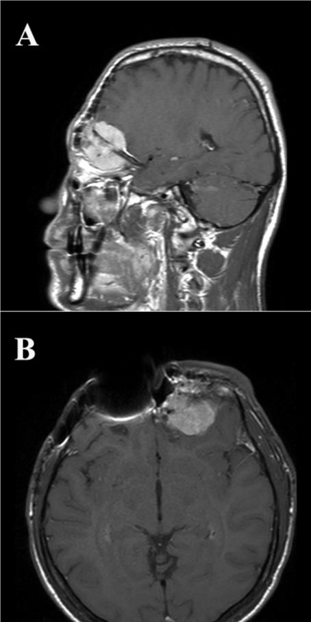
Figure 1:
Sagittal (A) and transverse (B) T1 MRI images post-gadolinium injection demonstrate a left-sided enhancing lesion infiltrating the ethmoidal-orbital region and the cerebral frontal lobe. Artifacts due to protesic material are present.
Read Full Article HTML DOI: 10.29328/journal.jnnd.1001039 Cite this Article Read Full Article PDF
More Images
Similar Articles
-
Direct Carotid Puncture for Flow Diverter Stent InsertionBhogal P*,Phillips TJ, Makalanda HLD. Direct Carotid Puncture for Flow Diverter Stent Insertion. . 2017 doi: 10.29328/journal.jnnd.1001004; 1: 024-028
-
Brain and immune system: KURU disease a toxicological process?Luisetto M*,Behzad Nili-Ahmadabadi,Ghulam Rasool Mashori,Ahmed Yesvi,Ram Kumar Sahu,Heba Nasser,Cabianca luca,Farhan Ahmad Khan. Brain and immune system: KURU disease a toxicological process?. . 2018 doi: 10.29328/journal.jnnd.1001010; 2: 014-027
-
Cranioplasty with preoperatively customized Polymethyl-methacrylate by using 3-Dimensional Printed Polyethylene Terephthalate Glycol MoldMehmet Beşir Sürme*,Omer Batu Hergunsel,Bekir Akgun,Metin Kaplan. Cranioplasty with preoperatively customized Polymethyl-methacrylate by using 3-Dimensional Printed Polyethylene Terephthalate Glycol Mold. . 2018 doi: 10.29328/journal.jnnd.1001016; 2: 052-064
-
Mimicking multiple sclerosis - Ghost tumor that comes and goes in different parts of the brain without any treatmentLong Ching*,Ming Him Yuen,Tak Lap Poon,Fung Ching Cheung,Shun Hin Ting,Wing Chi Fong. Mimicking multiple sclerosis - Ghost tumor that comes and goes in different parts of the brain without any treatment. . 2019 doi: 10.29328/journal.jnnd.1001020; 3: 087-090
-
The turing machine theory for some spinal cord and brain condition, A toxicological - antidotic depurative approachMauro Luisetto*,Behzad Nili Ahmadabadi,Ahmed Yesvi Rafa,Ram Kumar Sahu,Luca Cabianca,Ghulam Rasool Mashori,Farhan Ahmad Khan. The turing machine theory for some spinal cord and brain condition, A toxicological - antidotic depurative approach. . 2019 doi: 10.29328/journal.jnnd.1001023; 3: 102-134
-
Immunohistochemical expression of Nestin as Cancer Stem Cell Marker in gliomasRasha Mokhtar Abdelkareem*,Afaf T Elnashar,Khaled Nasser Fadle,Eman MS Muhammad. Immunohistochemical expression of Nestin as Cancer Stem Cell Marker in gliomas. . 2019 doi: 10.29328/journal.jnnd.1001027; 3: 162-166
-
Central nervous system diseases associated with blood brain barrier breakdown - A Comprehensive update of existing literatureRajib Dutta*. Central nervous system diseases associated with blood brain barrier breakdown - A Comprehensive update of existing literature. . 2020 doi: 10.29328/journal.jnnd.1001035; 4: 053-062
-
Atlantoaxial subluxation in the pediatric patient: Case series and literature reviewCatherine A Mazzola*,Catherine Christie,Isabel A Snee,Hamail Iqbal. Atlantoaxial subluxation in the pediatric patient: Case series and literature review. . 2020 doi: 10.29328/journal.jnnd.1001037; 4: 069-074
-
Primary intracranial Hodgkin’s lymphoma after a blunt trauma: A case reportLuca Riccioni*,Anna Maria Cremonini,Manlio Gessaroli. Primary intracranial Hodgkin’s lymphoma after a blunt trauma: A case report. . 2020 doi: 10.29328/journal.jnnd.1001039; 4: 079-083
-
Impact of mandibular advancement device in quantitative electroencephalogram and sleep quality in mild to severe obstructive sleep apneaCuspineda-Bravo ER*,García- Menéndez M,Castro-Batista F,Barquín-García SM,Cadelo-Casado D,Rodríguez AJ,Sharkey KM. Impact of mandibular advancement device in quantitative electroencephalogram and sleep quality in mild to severe obstructive sleep apnea. . 2020 doi: 10.29328/journal.jnnd.1001041; 4: 088-098
Recently Viewed
-
The Need of Wider and Deeper Skin Biopsy in Verrucous Carcinoma of the SoleLuca Damiani*,Giuseppe Argenziano,Andrea Ronchi,Francesca Pagliuca,Emma Carraturo,Vincenzo Piccolo,Gabriella Brancaccio. The Need of Wider and Deeper Skin Biopsy in Verrucous Carcinoma of the Sole. Ann Dermatol Res. 2025: doi: 10.29328/journal.adr.1001036; 9: 005-007
-
Comparative Analysis of Water Wells and Tap Water: Case Study from Lebanon, Baalbeck RegionChaden Moussa Haidar, Ali Awad, Walaa Diab, Farah Kanj, Hassan Younes, Ali Yaacoub, Marwa Rammal, Alaa Hamze. Comparative Analysis of Water Wells and Tap Water: Case Study from Lebanon, Baalbeck Region. Insights Vet Sci. 2024: doi: 10.29328/journal.ivs.1001043; 8: 018-027
-
A Case Report of Hepatic Rupture Associated with Hellp SyndromeSena-Martins M*,Daher PGS,Tadini V,Loprete GB,Nakata T,Mantuan L,Stefani NR,Vissechi-Filho CR,Souza GN,Sakita M. A Case Report of Hepatic Rupture Associated with Hellp Syndrome. Clin J Obstet Gynecol. 2025: doi: 10.29328/journal.cjog.1001192; 8: 077-079
-
Forensic Analysis of WhatsApp: A Review of Techniques, Challenges, and Future DirectionsNishchal Soni*. Forensic Analysis of WhatsApp: A Review of Techniques, Challenges, and Future Directions. J Forensic Sci Res. 2024: doi: 10.29328/journal.jfsr.1001059; 8: 019-024
-
Impact of Latex Sensitization on Asthma and Rhinitis Progression: A Study at Abidjan-Cocody University Hospital - Côte d’Ivoire (Progression of Asthma and Rhinitis related to Latex Sensitization)Dasse Sery Romuald*, KL Siransy, N Koffi, RO Yeboah, EK Nguessan, HA Adou, VP Goran-Kouacou, AU Assi, JY Seri, S Moussa, D Oura, CL Memel, H Koya, E Atoukoula. Impact of Latex Sensitization on Asthma and Rhinitis Progression: A Study at Abidjan-Cocody University Hospital - Côte d’Ivoire (Progression of Asthma and Rhinitis related to Latex Sensitization). Arch Asthma Allergy Immunol. 2024: doi: 10.29328/journal.aaai.1001035; 8: 007-012
Most Viewed
-
Feasibility study of magnetic sensing for detecting single-neuron action potentialsDenis Tonini,Kai Wu,Renata Saha,Jian-Ping Wang*. Feasibility study of magnetic sensing for detecting single-neuron action potentials. Ann Biomed Sci Eng. 2022 doi: 10.29328/journal.abse.1001018; 6: 019-029
-
Evaluation of In vitro and Ex vivo Models for Studying the Effectiveness of Vaginal Drug Systems in Controlling Microbe Infections: A Systematic ReviewMohammad Hossein Karami*, Majid Abdouss*, Mandana Karami. Evaluation of In vitro and Ex vivo Models for Studying the Effectiveness of Vaginal Drug Systems in Controlling Microbe Infections: A Systematic Review. Clin J Obstet Gynecol. 2023 doi: 10.29328/journal.cjog.1001151; 6: 201-215
-
Causal Link between Human Blood Metabolites and Asthma: An Investigation Using Mendelian RandomizationYong-Qing Zhu, Xiao-Yan Meng, Jing-Hua Yang*. Causal Link between Human Blood Metabolites and Asthma: An Investigation Using Mendelian Randomization. Arch Asthma Allergy Immunol. 2023 doi: 10.29328/journal.aaai.1001032; 7: 012-022
-
An algorithm to safely manage oral food challenge in an office-based setting for children with multiple food allergiesNathalie Cottel,Aïcha Dieme,Véronique Orcel,Yannick Chantran,Mélisande Bourgoin-Heck,Jocelyne Just. An algorithm to safely manage oral food challenge in an office-based setting for children with multiple food allergies. Arch Asthma Allergy Immunol. 2021 doi: 10.29328/journal.aaai.1001027; 5: 030-037
-
Postpartum as the best time for physical recovery and health careShizuka Torashima*,Mina Samukawa,Kazumi Tsujino,Yumi Sawada. Postpartum as the best time for physical recovery and health care. J Nov Physiother Rehabil. 2023 doi: 10.29328/journal.jnpr.1001049; 7: 001-007

If you are already a member of our network and need to keep track of any developments regarding a question you have already submitted, click "take me to my Query."


